Motivation
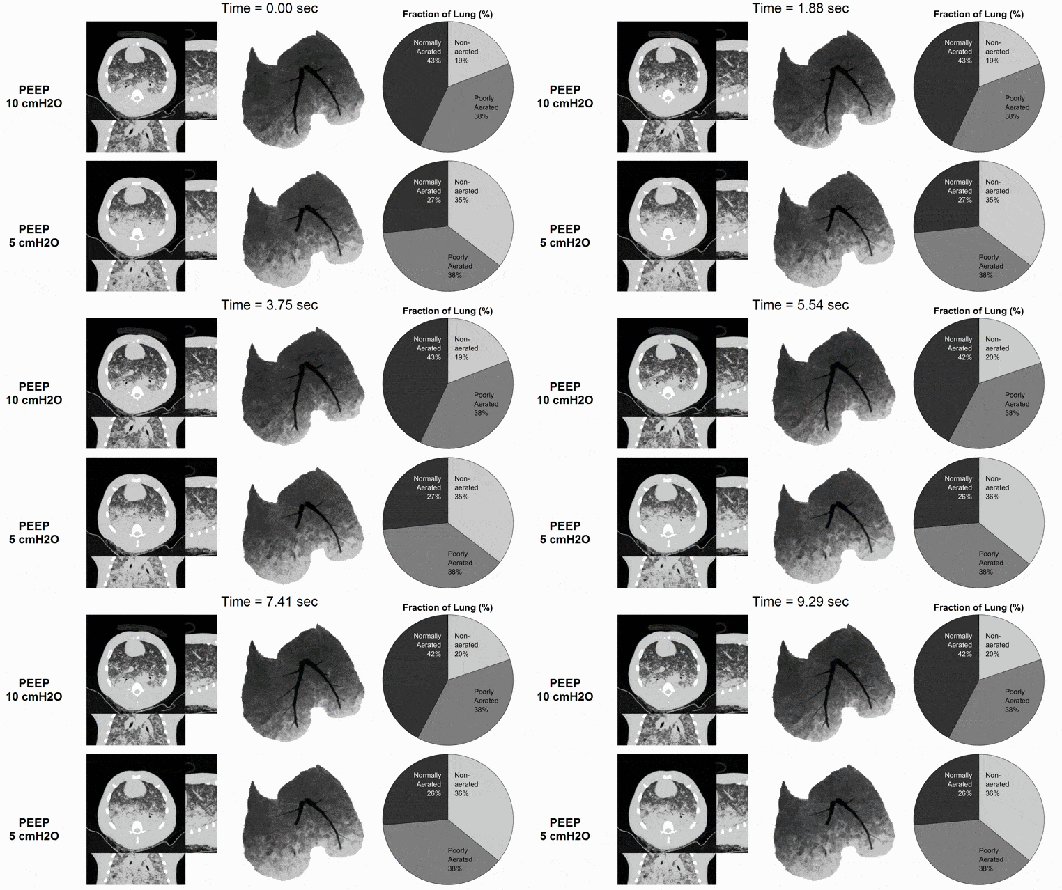
At the bedside, it is often difficult to infer the extent of lung injury and the severity of ventilation heterogeneity — but these factors influence a patient's response to changes in ventilation strategy (e.g., positive end-expiratory pressure or PEEP).
Our lab seeks to develop innovative approaches to dynamic medical imaging of pulmonary structure and function. We generate unique datasets that accurately capture how different regions of the injured lung respond to mechanical ventilator settings, without interrupting ventilation to take measurements.
Our goal is to quantitatively evaluate the safety and efficacy of mechanical ventilation strategies in experimental models of acute lung injury, and use these research findings to inform clinical practice.

We specialize in the acquisition, reconstruction, and analysis of dynamic X-ray computed tomography (CT) scans of the lungs, capturing periodic motion during uninterrupted breathing or ventilation.

Dynamic CT Imaging
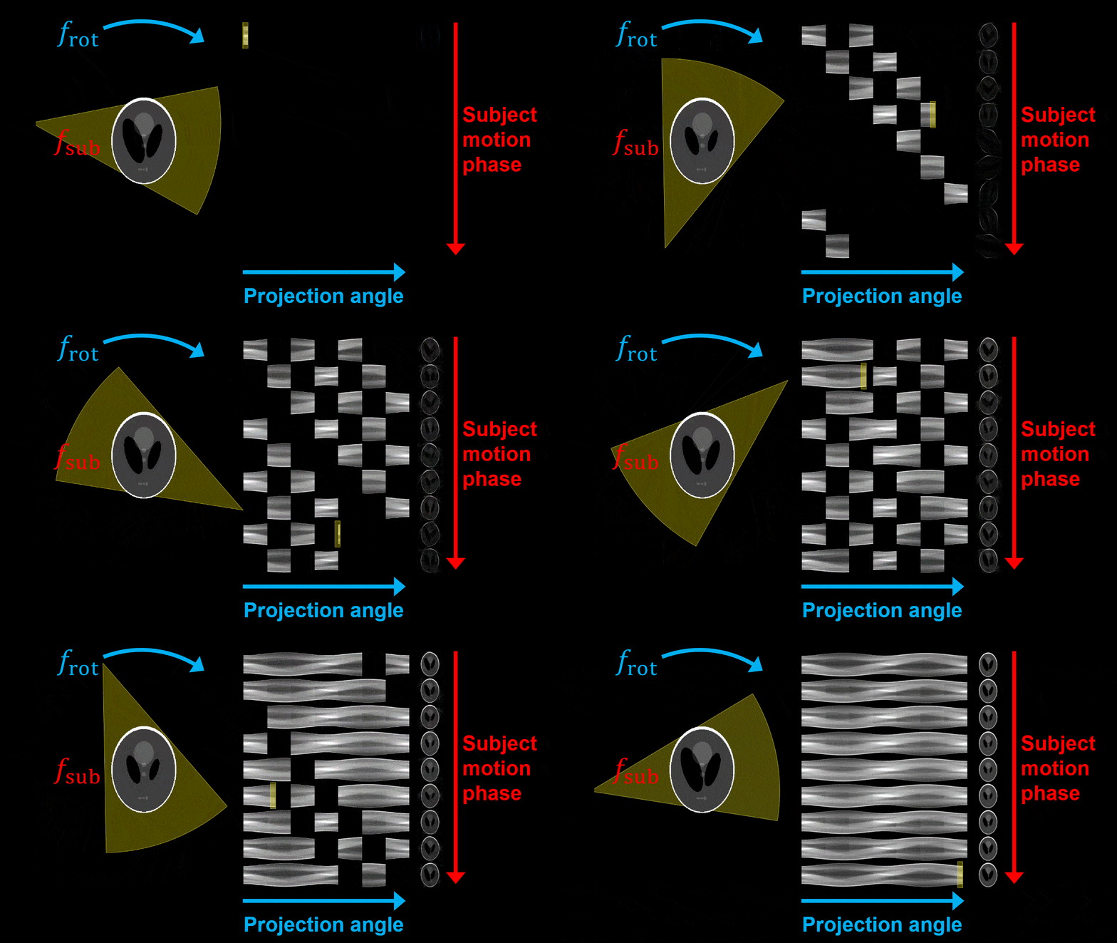
X-ray projections are acquired continuously while the scanner rotates the X-ray source and detector around the subject at a frequency frot.
Simultaneously, the subject exhibits periodic motion at a frequency fsub.
Each projection is sorted according to both scanner angle and subject motion phase, allowing reconstruction of a sequence of images representing the periodic subject motion.
Learn more: Go to journal site

Image Registration
Registration is the process of estimating a spatial transformation (arrows) that aligns the structures of a moving image or source image (red) with the structures of a fixed image or target image (blue).
We apply cyclic groupwise registration algorithms to our dynamic CT image sequences. This allows us to quantitatively analyze the periodic motion of lung tissue throughout the ventilation cycle.
Learn more: Go to journal site

Dynamic Strain Measurement
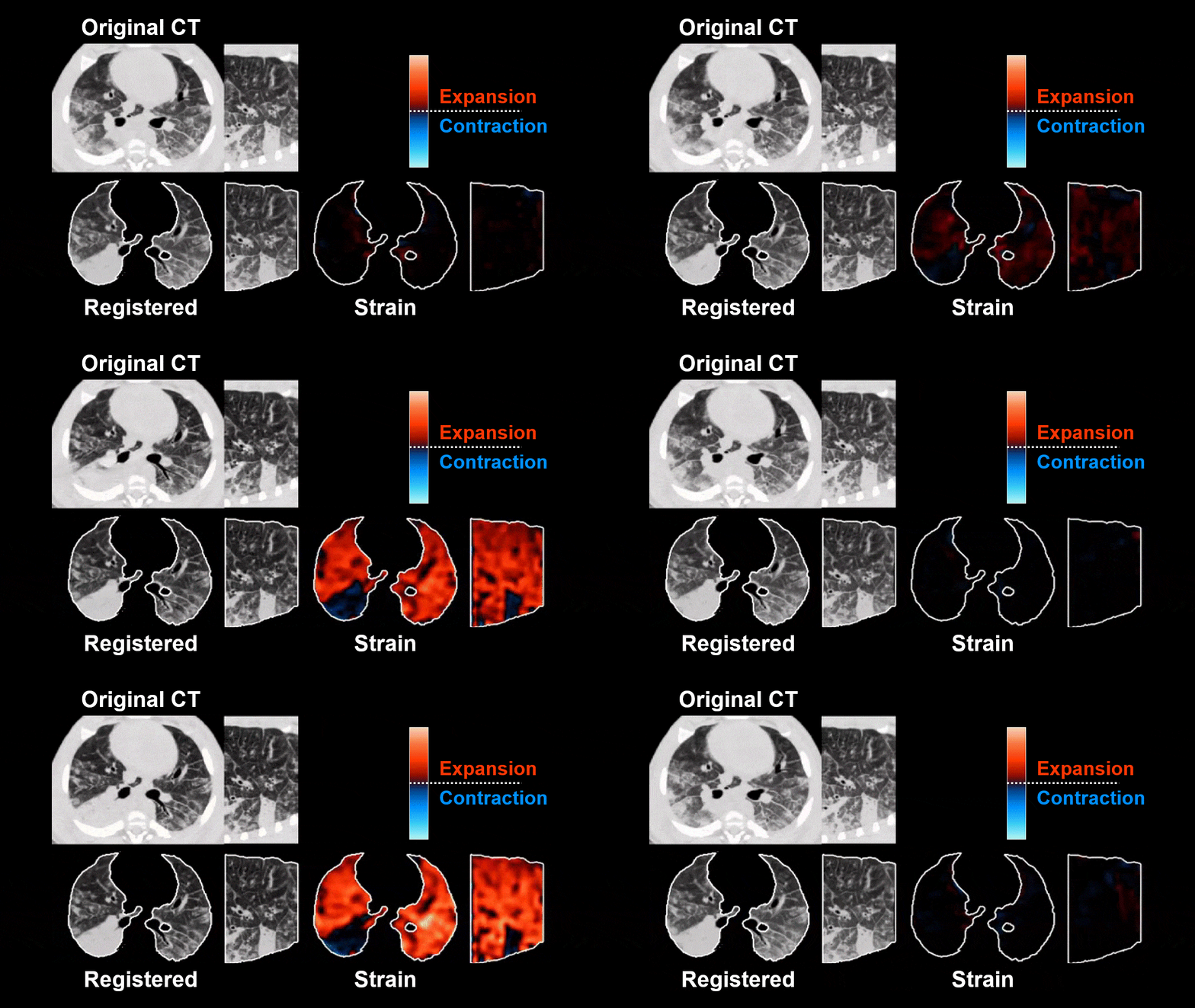
Registered image sequences should appear relatively motionless, with time-varying intensity reflecting changes in regional aeration status during the ventilation cycle.
Regional strain or volume change is estimated as the "Jacobian" or the determinant of the deformation gradient, using the spatial transformation estimated by registration.
Learn more: Go to journal site

Dynamic Aeration Measurement
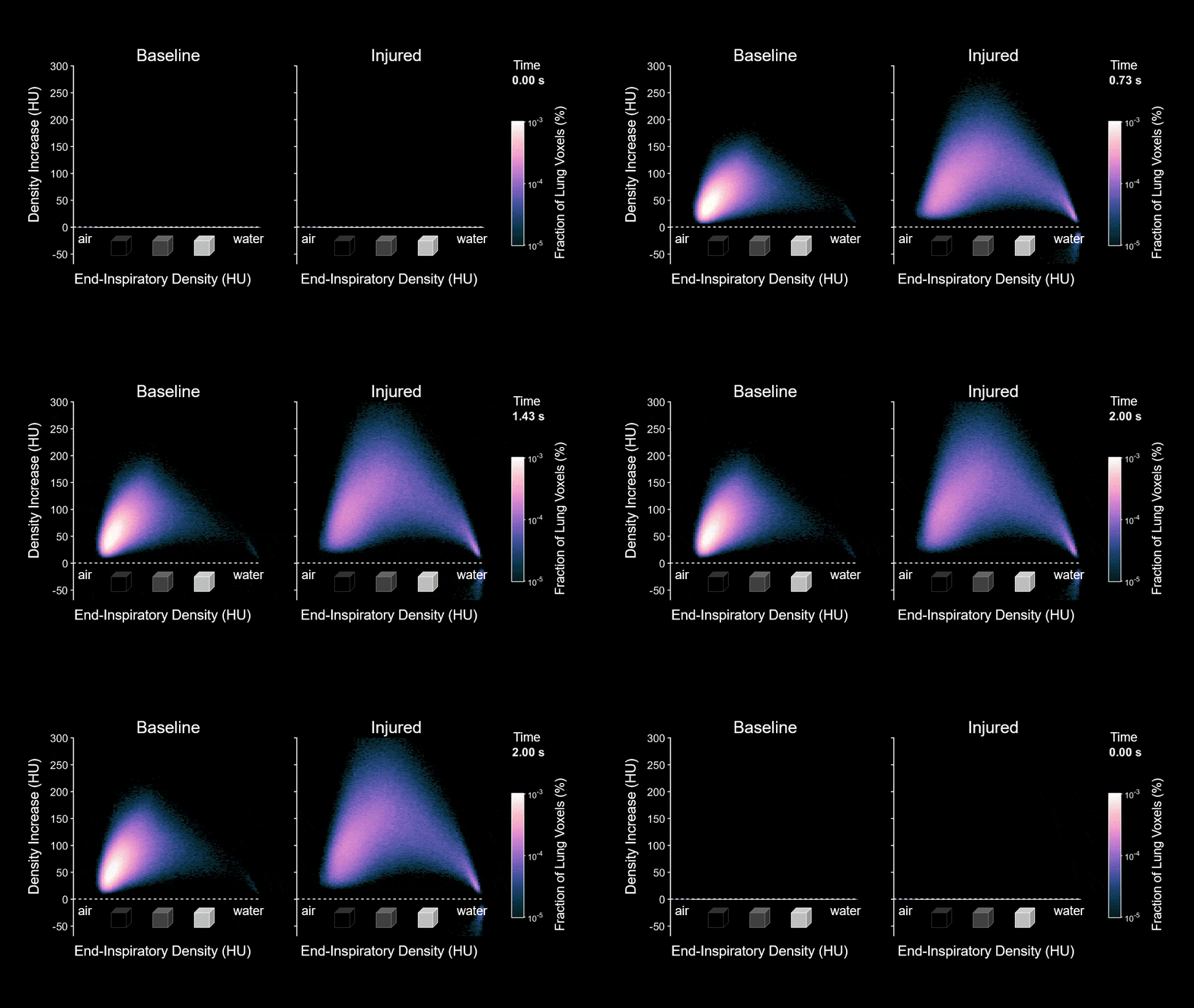
Rapid loss of regional aeration during exhalation may indicate the underlying process of microscale alveolar collapse or derecruitment, an important factor in ventilator-induced lung injury.
Imaging-based measurements demonstrate larger, faster loss of aeration in poorly aerated regions of the injured lung. These findings provide support for alternative modalities of mechanical ventilation that rely on reduced exhalation time to avoid cyclic alveolar collapse.
Learn more: Go to journal site