Motivation
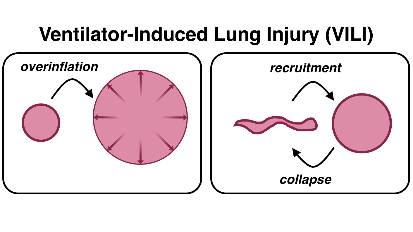
Mechanical ventilation saves lives — and it has the potential to cause serious harm by damaging alveoli, often in ways that are difficult to observe at the bedside.
Our lab seeks to identify injurious processes occurring dynamically in unstable alveoli, connect these microscale phenomena to regional dynamics observed in medical imaging, and ultimately develop mechanical ventilation strategies that avoid these harmful processes.
We use dynamic microscopic imaging techniques with respiratory-gated reconstruction to reveal how alveolar structures move and deform during uninterrupted ventilation.
Dynamic Microscopic Imaging
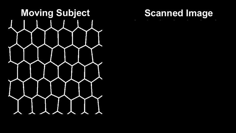
Laser-scanning confocal microscopy produces micron-resolution images using a raster pattern, which results in severe distortions if the subject is moving.
We acquire and retrospectively sort the scanned rows according to the corresponding phase of the ventilation cycle, producing an image sequence which describes the periodic subject motion.
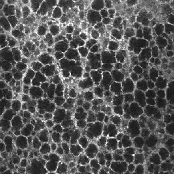
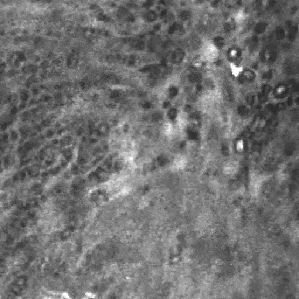
Normally aerated regions exhibit relatively uniform deformations of alveoli:
Poorly aerated regions exhibit cyclic collapse and reopening of damaged alveoli:
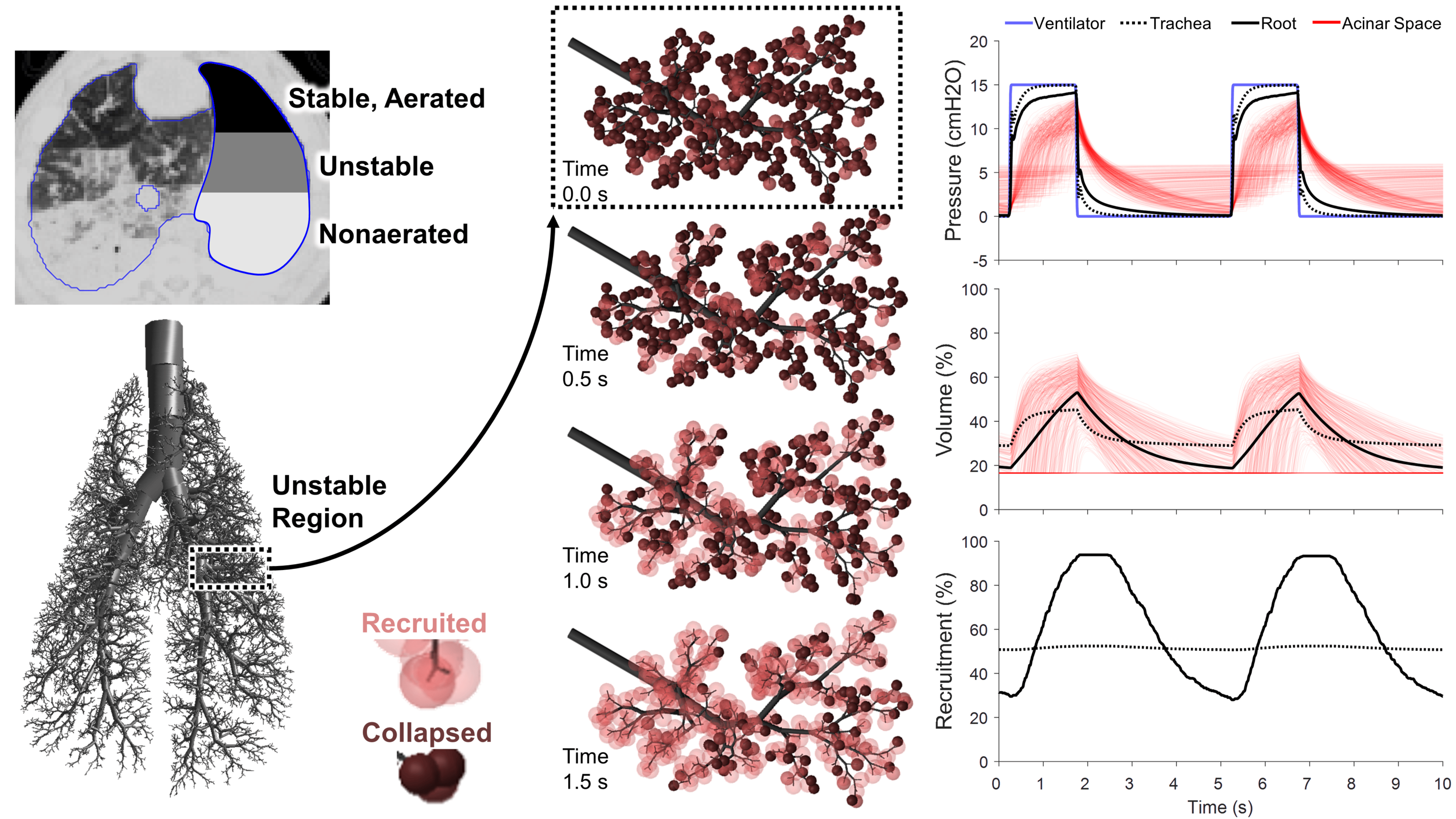
Computational Modeling
Our experimental observations guide our computational modeling efforts, which allow us to test a much wider range of pathophysiologic conditions.
Computational simulations yield new hypotheses about how alveolar inflation instability occurs, and how we might prevent intratidal alveolar collapse using novel ventilation modalities.